File:Anterior chamber angle.jpg

Original file (1,365 × 1,096 pixels, file size: 336 KB, MIME type: image/jpeg)
Summary
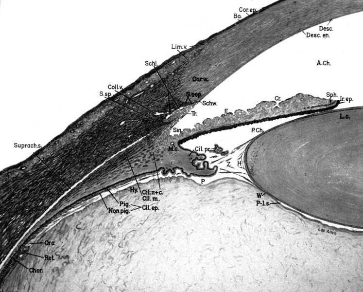
Anatomy of the anterior chamber angle.
Sketch of the anterior chamber angle. The labeled structures (listed alphabetically) are: A. Ch, anterior chamber; Bo., Bowman’s layer; Chor, choroid; Cil. ep., ciliary epithelium; Cil. m., ciliary muscle (longitudinal); Cil. pr., ciliary process; Cil. r. + c., ciliary body (radial and circular muscles); Coll. v., collector veins; Cor. ep., corneal epithelium; Cor. w., corneal wedge; Cr., iris crypt; Desc, Descemet’s membrane; Desc. en., corneal endothelium (or Descemet’s endothelium); F,iris furrow; H, Hanover’s canal; Hy., hyaloid; Ir. ep, iris pigment epithelium; c.,lens cortex; Lim. v., limbal vessels; M. c., major circle of iris; Non pig, nonpigmented ciliary epithelium; Ora, ora serrata; P, Petit’s canal; Pig., pigmented ciliary epithelium; P. Ch., posterior chamber; P-l. s., postlenticular space; Ret., retina; Schl., Schlemm’s canal; Schw., Schwalbe’s line; Sin., angle recess (or sinus); Sph., sphincter; S. sep., sclera septum; S. sp., scleral spur; Suprach. s., suprachoroidal space; Tr., trabecular meshwork; W, Wieger’s ligament; Z, zonules. (Because this sketch was drawn in the 1940s, some of the terms, such as Descemet’s endothelium, are different from those used today.)
Image from the Academy's Image Collection (copyright by the American Academy of Ophthalmology). https://www.aao.org/education/image/anterior-chamber-angle-17
File history
Click on a date/time to view the file as it appeared at that time.
| Date/Time | Thumbnail | Dimensions | User | Comment | |
|---|---|---|---|---|---|
| current | 13:44, May 31, 2026 | | 1,365 × 1,096 (336 KB) | Érico.Sant'Anna (talk | contribs) | Anatomy of the anterior chamber angle. Sketch of the anterior chamber angle. The labeled structures (listed alphabetically) are: A. Ch, anterior chamber; Bo., Bowman’s layer; Chor, choroid; Cil. ep., ciliary epithelium; Cil. m., ciliary muscle (longitudinal); Cil. pr., ciliary process; Cil. r. + c., ciliary body (radial and circular muscles); Coll. v., collector veins; Cor. ep., corneal epithelium; Cor. w., corneal wedge; Cr., iris crypt; Desc, Descemet’s membrane; Desc. en., corneal endothe... |
You cannot overwrite this file.
File usage
The following page uses this file: