

File:Visualization of Submacular SPCAs Through Progressive Geographic Atrophy.png
Original file (7,382 × 4,979 pixels, file size: 28.14 MB, MIME type: image/png)
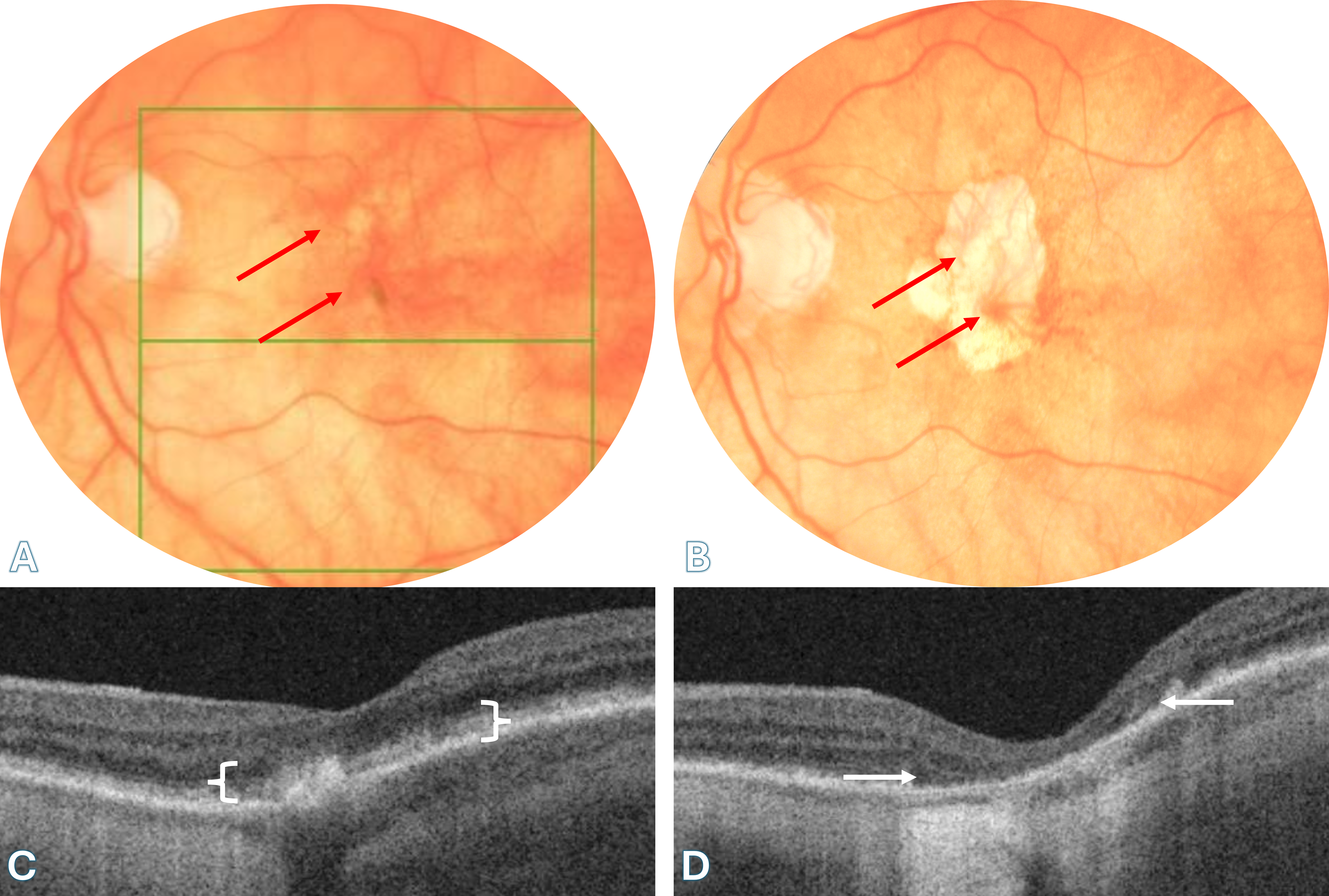
Fundus photographs (A, B) and OCT scans (C, D) illustrating the 5-year progression of geographic atrophy (GA) in an 85-year-old woman. (A) At baseline, subtle pigmentary changes are seen in the macula, with two early visible submacular vessels presumed to be short posterior ciliary arteries (SPCAs) (red arrows). (B) After five years, the GA has enlarged significantly, unmasking the underlying submacular choroidal vascular network due to loss of overlying retinal pigment epithelium (RPE) and photoreceptors. The two spoke-like SPCAs remain visible, marking the entry points where they penetrate the sclera to supply the submacular choroid (red arrows). These vessels are distinctly more narrowed than in the baseline image, likely reflecting vascular remodeling. They must be distinguished from posterior vortex vein ampullae. (C) OCT at baseline shows intact outer retina and preserved RPE over the fovea. (D) Follow-up OCT at the site of GA reveals complete outer retinal atrophy, including the photoreceptor layer and RPE, with resulting collapse of the inner retina (white arrows) toward the now-thinned choroid. The atrophic window allows direct visualization of choroidal structures, which correlate precisely with the fundus findings in panel B. (Courtesy of J. Khadamy)
File history
Click on a date/time to view the file as it appeared at that time.
| Date/Time | Thumbnail | Dimensions | User | Comment | |
|---|---|---|---|---|---|
| current | 03:10, June 24, 2025 | | 7,382 × 4,979 (28.14 MB) | Joobin.Khadamy (talk | contribs) |
You cannot overwrite this file.
File usage
The following page uses this file:





