

Congenital Retinal Macrovessel
All content on Eyewiki is protected by copyright law and the Terms of Service. This content may not be reproduced, copied, or put into any artificial intelligence program, including large language and generative AI models, without permission from the Academy.
Disease Entity
Congenital retinal macrovessel (CRM) is a rare congenital retinal vascular anomaly characterized by an enlarged, aberrant retinal vessel—most often venous—that traverses the macular region and may cross the horizontal raphe. The lesion was first described by Mauthner[1]. Archer et al. subsequently classified similar lesions within their spectrum of retinal arteriovenous communications, corresponding to group 1 in their classification system[2].
The term congenital retinal macrovessel was introduced by Brown et al. after their report of seven patients with an anomalous retinal vessel involving the macula[3]. More recently, Pichi et al. proposed the term retinal venous malformation (RVM), based on the predominantly venous origin of these lesions and their reported association with cerebral venous malformations[4]. Although both terms are used in the literature, CRM remains widely recognized in clinical practice.
Disease
CRM typically consists of a large, dilated, anomalous retinal vessel that crosses the macular region and supplies or drains retinal territories on both sides of the horizontal raphe. The vessel may traverse the fovea, distort the foveal avascular zone (FAZ), communicate with perifoveal capillary networks, or be associated with abnormal branching of the temporal retinal vasculature.
Most lesions appear to be of venous origin; arterial variants are rare. In the multicenter series reported by Pichi et al., only four lesions were considered potentially arterial; however, fluorescein angiography was not performed in all of these cases, limiting definitive confirmation of vessel type[4].
Population-based epidemiologic data are limited. A prevalence of approximately 1 in 200,000 has been cited; however, the true prevalence is unknown and may be underestimated because many lesions are asymptomatic and detected incidentally during routine fundus examination. In the largest multicenter case series published to date, 49 cases were identified, all of which were unilateral. Patients were predominantly female (65%), and the mean age at diagnosis was 44 years[4]. The condition may be identified at any age, particularly when a dilated fundus examination or retinal imaging is performed for unrelated reasons.
Etiology
CRM is presumed to result from abnormal retinal vascular development during embryogenesis. The proposed timing corresponds to the developmental period during which the retinal vascular network and macular vascular architecture are established[2].
Diagnosis

The diagnosis of CRM is primarily based on funduscopic examination, which usually allows direct visualization of a large, aberrant vessel traversing the macular region and crossing the horizontal raphe. The anomalous vessel may cross or skirt the fovea, alter the FAZ, and communicate with perifoveal vascular networks. In the largest available series, RVMs were most commonly located in the inferotemporal quadrant (60%), followed by the superotemporal quadrant (33%) and the papillomacular bundle (approximately 10%)[4].
Signs
Associated funduscopic features may include:
- Abnormal branching of the temporal retinal veins.
- Perifoveal vascular loops or small arteriovenous communications.
- An indistinct, displaced, or absent visible FAZ.
- Localized retinal pigment epithelium alterations.
- Preretinal or intraretinal hemorrhage, exudation or macular edema in complicated cases.
- Collateral vessels or signs of previous retinal vascular occlusion[5].
Symptoms
Most affected individuals are asymptomatic and retain good visual acuity. When symptoms occur, they are usually related to foveal involvement, associated foveal developmental abnormalities, or secondary retinal complications. Symptoms may include blurred vision, reduced central visual acuity, metamorphopsia, or central or paracentral scotoma.
Diagnostic Procedures
Multimodal imaging is valuable for defining the vessel’s course, caliber, depth, relationship to the fovea, and associated macular abnormalities.
Optical Coherence Tomography
Structural optical coherence tomography (OCT), including spectral-domain OCT and swept-source OCT, is useful for assessing foveal anatomy and associated retinal complications. Findings may include:
- A hyperreflective intraretinal vascular lumen with posterior shadowing.
- Distortion of the foveal contour.
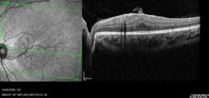
The near-infrared reflectance image (left) shows an anomalous vessel traversing the macular region. The corresponding OCT B-scan (right) demonstrates a hyperreflective intraretinal vascular profile with posterior shadowing and associated foveal hypoplasia. - Intraretinal cysts or cystoid macular edema.
- Subretinal fluid or serous retinal detachment[6].
- Foveal thinning, disorganization, or structural changes associated with macular ischemia.
- Persistence of inner retinal layers at the fovea or other features of foveal hypoplasia.
- Epiretinal membrane or vitreomacular interface abnormalities, when present.

Optical Coherence Tomography Angiography
Optical coherence tomography angiography (OCTA) noninvasively delineates the macrovessel and defines its relationship to the superficial, intermediate, and deep retinal capillary plexuses. OCTA may demonstrate FAZ distortion or absence, abnormal vascular connections, focal capillary rarefaction, and areas of nonperfusion.
In some cases, OCTA shows increased flow signal within the macrovessel relative to adjacent retinal vessels and anomalous vascular communications between retinal arteries and the venous macrovessel. Abnormalities may be particularly conspicuous in the intermediate and deep capillary plexuses. OCTA is also useful for longitudinal assessment of macular complications and for excluding secondary macular neovascularization[7][8].
Fluorescein Angiography
Fluorescein angiography may show early filling and delayed emptying of the anomalous vessel. A dilated perifoveal capillary network may be present. Additional findings can include focal capillary dilatation, areas of capillary nonperfusion, perifoveal arteriovenous communications, and blocked fluorescence caused by overlying preretinal hemorrhage[9].
Leakage is generally absent in uncomplicated CRM. The presence of leakage should prompt evaluation for secondary macular edema, macroaneurysm, retinal vascular occlusion, macular neovascularization, or another coexisting retinal disorder.
Color Fundus Photography
Color fundus photography provides baseline documentation of the macrovessel’s morphology, its relationship to the fovea and optic disc, and associated hemorrhagic or exudative changes. Serial photography may be useful for documenting stability over time.
Differential Diagnosis
- Retinal arteriovenous malformation is a congenital vascular anomaly characterized by direct arteriovenous communication without an intervening capillary bed. The clinical spectrum ranges from small, isolated lesions to markedly dilated and tortuous vascular networks with a characteristic “bag-of-worms” appearance. Extensive lesions are often associated with Wyburn–Mason syndrome, which is characterized by retinal and intracranial arteriovenous malformations, particularly involving the midbrain, and, less commonly, facial vascular malformations. Ocular Manifestations of Wyburn-Mason Syndrome Jump to:navigation, search
- Retinal capillary hemangioblastoma: circumscribed retinal vascular tumor with prominent feeding and draining vessels, exudation, and potential association with von Hippel–Lindau disease. Retinal Capillary Hemangioblastoma and von Hippel-Lindau Disease Jump to:navigation, search
- Retinal cavernous hemangioma: dark-red, grape-like cluster of thin-walled saccular aneurysms; fluorescein angiography typically demonstrates slow, incomplete filling, plasma–erythrocyte layering (“fluorescein capping”), and minimal or absent leakage.
- Familial retinal arteriolar tortuosity: bilateral arteriolar tortuosity, sometimes associated with recurrent retinal hemorrhage; does not usually produce a solitary macular venous macrovessel.
- Choroidal vascular anomalies, particularly choroidal macrovessels, which appear funduscopically as large, tortuous, orange-red vessels, sometimes accompanied by retinal pigment epithelial changes. OCT confirms their choroidal location, demonstrating an enlarged hyporeflective vascular channel with possible elevation of the overlying outer retina.Choroidal Vascular Anomalies: Posterior Vortex Veins, Choroidal Macrovessels, Choroidal Macroaneurysms, and Choroidal Vessel Varix
Systemic Associations and Evaluation
The most important reported systemic association is with cerebral vascular malformations, particularly venous malformations. In the multicenter cohort reported by Pichi et al., cerebral venous malformations were identified in approximately 24% of patients with CRM, exceeding the reported prevalence of 0.2%–6.0% in the general population. Cerebral cavernous malformations were identified in 4 of 49 patients (8%). Cerebral vascular anomaly was ipsilateral to the retinal lesion in most cases (83%) and frequently involved the frontal lobe, observed in 75% of cases[4].
These findings support a possible developmental relationship between retinal venous malformations and cerebral venous malformations. However, the evidence is derived primarily from retrospective series, and the true prevalence of cerebral abnormalities among all patients with CRM remains uncertain.
Brain MRI with contrast should therefore be considered, particularly in patients with newly diagnosed CRM, atypical retinal features, neurological symptoms, a family history of vascular malformations, or no prior neuroimaging. Neuroimaging decisions should be individualized in collaboration with neurology, neuroradiology, as appropriate.
Management
General Treatment
Evidence guiding the management of CRM is derived primarily from case reports and small case series. Uncomplicated CRM are generally managed conservatively, with intervention reserved for associated vision-threatening complications rather than the anomalous vessel itself. Baseline assessment should include best-corrected visual acuity, dilated fundus examination, color fundus photography, and structural OCT, supplemented by OCTA when available.
Macular edema warrants careful characterization, as its mechanism may not always be exudative. Intraretinal cystic changes may occur without fluorescein leakage, potentially reflecting altered local hemodynamics or increased venous hydrostatic pressure rather than blood–retinal barrier disruption. Observation may be appropriate when edema is mild, visual acuity remains stable, fluorescein angiography shows no leakage and OCTA excludes macular neovascularization, as spontaneous resolution has been reported[10][11].
Persistent or progressive edema, hemorrhage, exudation, angiographic leakage, or declining visual acuity should prompt evaluation for treatable associated conditions, including retinal arterial macroaneurysm, retinal vein occlusion, macular neovascularization, vitreomacular interface abnormalities, and Valsalva retinopathy[5]. Treatment should target the underlying mechanism and may include intravitreal anti-VEGF agents[12][13] or focal laser photocoagulation[14] when appropriate. Published evidence supporting oral corticosteroids or intravitreal corticosteroid implants for macular edema directly attributable to congenital retinal macrovessels remains lacking.
Medical Follow-up
Follow-up intervals should be individualized according to symptoms, foveal involvement, OCT findings, and the presence of complications. Patients should be advised to seek prompt ophthalmic assessment for new metamorphopsia, central scotoma, acute visual decline or new floaters.
Prognosis
CRM generally has a favorable visual prognosis. The lesion is typically unilateral, stable, and nonprogressive, and most affected individuals remain asymptomatic with preserved visual acuity[15]. In the largest multicenter series, 80% had no ocular complications, and the mean best-corrected visual acuity was 20/25[4].
Foveal crossing by the macrovessel does not necessarily predict poor visual acuity. In uncomplicated eyes, visual acuity was not significantly different between lesions that crossed the fovea and those that did not[16]. Visual impairment is more strongly associated with foveal structural abnormalities and secondary retinal complications than with the presence of the macrovessel alone.
OCT features associated with reduced visual acuity may include an absent or shallow foveal pit, persistence of inner retinal layers at the fovea, foveal cystic change, outer nuclear layer widening, ellipsoid-zone disruption, and macular edema. CRM-associated foveal hypoplasia may result in stable long-term visual impairment, reflecting altered foveal development rather than progressive injury caused by the anomalous vessel[17].
In the largest available series, ocular complications occurred in 20% and were associated with lower mean visual acuity. Reported complications include macular edema, retinal hemorrhage, deep capillary plexus aneurysmal changes, retinal arterial macroaneurysm, retinal vascular occlusion, macular ischemia, serous macular detachment, and macular neovascularization[4]. A causal association between CRM and certain complications remains uncertain, as some may be coincidental rather than directly attributable to the macrovessel.
References
- ↑ Mauthner L. Lehrbuch der Ophthalmoscopie. Vienna, Austria: Tendler Co; 1869:249.
- ↑ 2.0 2.1 Brown GC, Donoso LA, Magargal LE, Goldberg RE, Sarin LK. Congenital retinal macrovessels. Arch Ophthalmol. 1982;100(9):1430-1436.
- ↑ Archer, D. B., Deutman, A., Ernest, J. T., & Krill, A. E. (1973). Arteriovenous communications of the retina. American journal of ophthalmology, 75(2), 224–241.
- ↑ 4.0 4.1 4.2 4.3 4.4 4.5 4.6 Pichi, F., Freund, K. B., Ciardella, A., Morara, M., Abboud, E. B., Ghazi, N., Dackiw, C., Choudhry, N., Souza, E. C., Cunha, L. P., Arevalo, J. F., Liu, T. Y. A., Wenick, A., He, L., Villarreal, G., Jr, Neri, P., & Sarraf, D. (2018). Congenital Retinal Macrovessel and the Association of Retinal Venous Malformations With Venous Malformations of the Brain. JAMA ophthalmology, 136(4), 372–379.
- ↑ 5.0 5.1 de Crecchio, G., Alfieri, M.C., Cennamo, G. et al. Congenital macular macrovessels. Graefe's Arch Clin Exp Ophthalmo 244, 1183–1187 (2006). https://doi.org/10.1007/s00417-005-0213-2
- ↑ Kumar V, Ghosh B, Raina U, Goel N. Central serous chorioretinopathy in a patient with congenital retinal macrovessel. Can J Ophthalmol 2009;44:e57
- ↑ Preziosa, C., Milani, P., Ciasca, P., Bergamini, F., Staurenghi, G., & Pellegrini, M. (2021). OPTICAL COHERENCE TOMOGRAPHY ANGIOGRAPHY FINDINGS IN A CASE OF CONGENITAL RETINAL MACROVESSEL WITH ANOMALOUS RETINAL ANASTOMOSIS ASSOCIATED WITH CONTRALATERAL MYELINATED NERVE FIBERS AND RETINAL VASCULAR ABNORMALITIES. Retinal cases & brief reports, 15(5), 605–610.
- ↑ Chawla, R., Bypareddy, R., Tripathy, K., Daggumili, S. S., & Tomar, A. S. (2016). Optical Coherence Tomography Angiography Imaging of Congenital Retinal Macrovessel. Ophthalmic surgery, lasers & imaging retina, 47(10), 972–973. https://doi.org/10.3928/23258160-20161004-14
- ↑ Petropoulos, I. K., Petkou, D., Theoulakis, P. E., Kordelou, A., Pournaras, C. J., & Katsimpris, J. M. (2008). Congenital retinal macrovessels: description of three cases and review of the literature. Klinische Monatsblatter fur Augenheilkunde, 225(5), 469–472.
- ↑ Beatty S, Goodall K, Radford R et al. Decompensation of a congenital retinal macrovessel with arteriovenous communications induced by repetitive rollercoaster rides. Am J Ophthalmol 2000; 130: 527–528.
- ↑ Zheng, F., Lai, K., Yin, H., He, J., Xu, Y., & Ye, P. (2022). Case Report: Self-Resolved Macular Edema Secondary to Congenital Retinal Macrovessels. Frontiers in medicine, 8, 771007.
- ↑ Goel, N., Kumar, V., Seth, A., & Ghosh, B. (2015). Intravitreal bevacizumab in congenital retinal macrovessel with retinal arteriolar macroaneurysm. Saudi journal of ophthalmology : official journal of the Saudi Ophthalmological Society, 29(4), 292–294.
- ↑ Al-Zamil, W. M., & Magliyah, M. S. (2020). Favourable outcome in retinal macrovessel-related choroidal neovascularization. Saudi journal of ophthalmology : official journal of the Saudi Ophthalmological Society, 34(2), 139–141.
- ↑ Chalam KV, Gupta SK, Vinjamaram S, Shah VA. Congenital Anomalous Retinal Artery Associated With a Leaking Macroaneurysm. Arch Ophthalmol. 2003;121(3):409–410.
- ↑ Crecchio, G.d., Pacente, L., Alfieri, M.C., Pignalosa, G. and Greco, G.M. (1999), Congenital retinal macrovessels, A "low visual acuity" case report with a 14-year follow-up. Acta Ophthalmologica Scandinavica, 77: 474-475.
- ↑ Shah, V., Reddy, M. A., & Papastefanou, V. P. (2017). Multimodal Imaging Analysis in a Case with Congenital Fovea-Involving Retinal Macrovessel and Excellent Visual Acuity. Case reports in ophthalmological medicine, 2017, 4057615.
- ↑ Kohli, P., Mishra, C., Baliga, G., & Rajan, R. P. (2022). Multimodal imaging of congenital retinal macrovessel with secondary foveal hypoplasia. BMJ case reports, 15(4), e249563.

