File:Aos17588-fig-0001-m.jpg
Original file (2,128 × 3,778 pixels, file size: 969 KB, MIME type: image/jpeg)
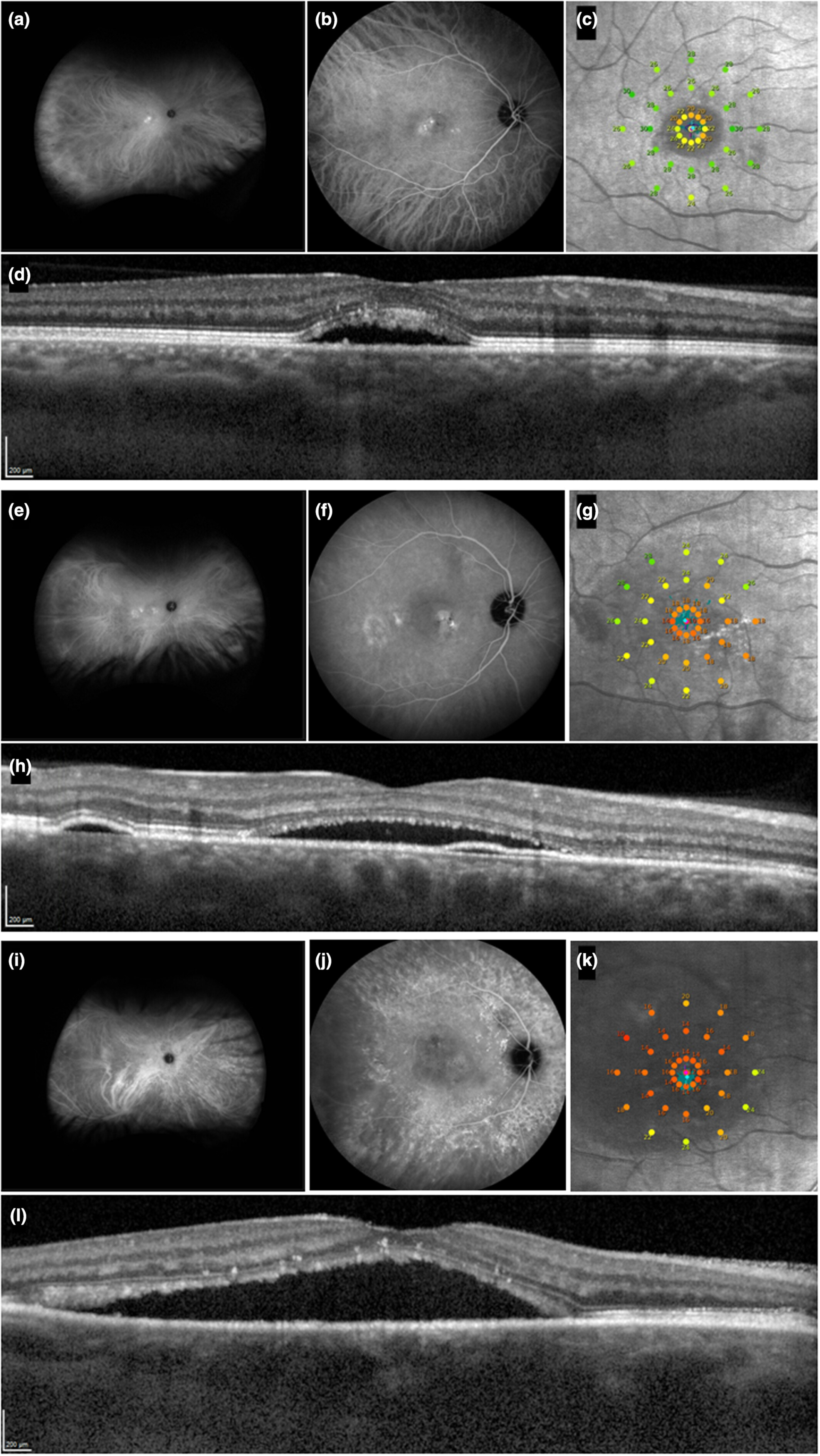
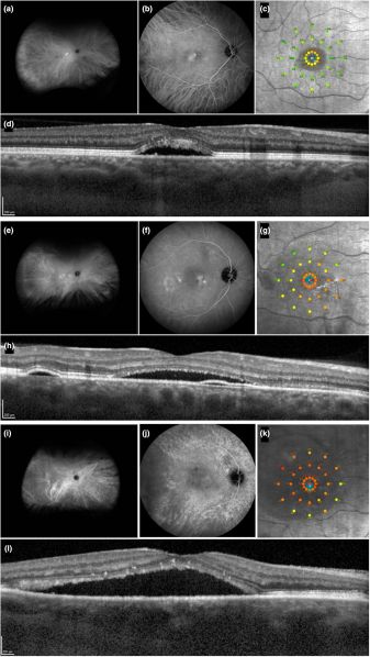
FIGURE 1. Ultra-widefield indocyanine green angiography (ICGA) images (a, e, i), 55° ICGA images (b, f, j), microperimetry (c, g, k), and optical coherence tomography (d, h, l) results of patients with central serous chorioretinopathy showing unifocal signs of indistinct choroidal hyperpermeability (uni-FISH) (a–d), multifocal signs of indistinct choroidal hyperpermeability (multi-FISH) (e–h), and diffuse signs of indistinct choroidal hyperpermeability (DISH) (i–l). Note the colocalisation of lower sensitivity with hyperpermeability in uni-FISH and multi-FISH, as well as the relatively lower sensitivity in DISH.
File history
Click on a date/time to view the file as it appeared at that time.
| Date/Time | Thumbnail | Dimensions | User | Comment | |
|---|---|---|---|---|---|
| current | 08:18, February 4, 2026 | | 2,128 × 3,778 (969 KB) | Camiel.Boon (talk | contribs) |
You cannot overwrite this file.
File usage
The following page uses this file: