File:Multimodal Imaging Demonstrating Pachyvessels in Pachychoroid Spectrum Disorder.jpg
Original file (2,369 × 3,978 pixels, file size: 1.43 MB, MIME type: image/jpeg)
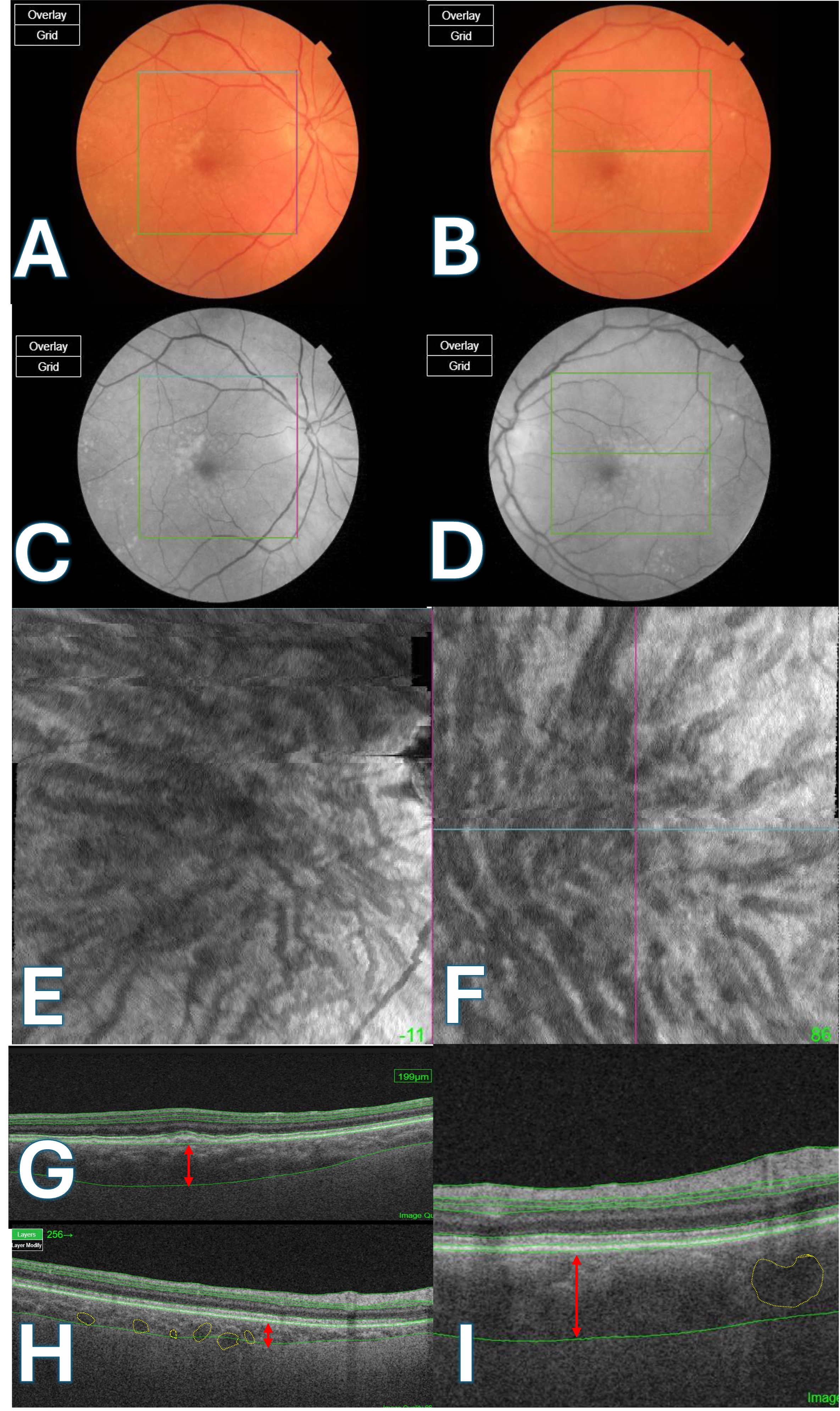
(A, B) Color fundus photographs of the right and left eyes, respectively. Note the presence of subtle macular pigmentary changes.
(C, D) Red-free images of the right and left eyes, respectively. These images enhance the visibility of retinal vasculature and subtle retinal changes.
(E, F) En-face OCT images at the choroid level show dilated choroidal vessels (Pachyvessels).
(G) Enhanced Depth Imaging Optical Coherence Tomography (EDI-OCT) of the right eye demonstrated increased choroidal thickness (red arrow), particularly in the superior part of the macula.
(H) EDI-OCT of the right eye with binarization highlighting large choroidal vessels (Pachyvessels outlined in yellow). The red arrow shows the choroidal thickness measurement in the inferior macula.
(I) EDI-OCT of the left eye showing a thickened choroid (red arrow), and dilated choroidal vessels (Pachyvessels outlined in yellow). Overall, the left eye exhibits more prominent pachyvessels and a generally thicker choroid compared to the right eye. (Image courtesy of J. Khadamy)
File history
Click on a date/time to view the file as it appeared at that time.
| Date/Time | Thumbnail | Dimensions | User | Comment | |
|---|---|---|---|---|---|
| current | 08:41, March 1, 2025 | | 2,369 × 3,978 (1.43 MB) | Joobin.Khadamy (talk | contribs) | arrows |
| 08:16, March 1, 2025 | 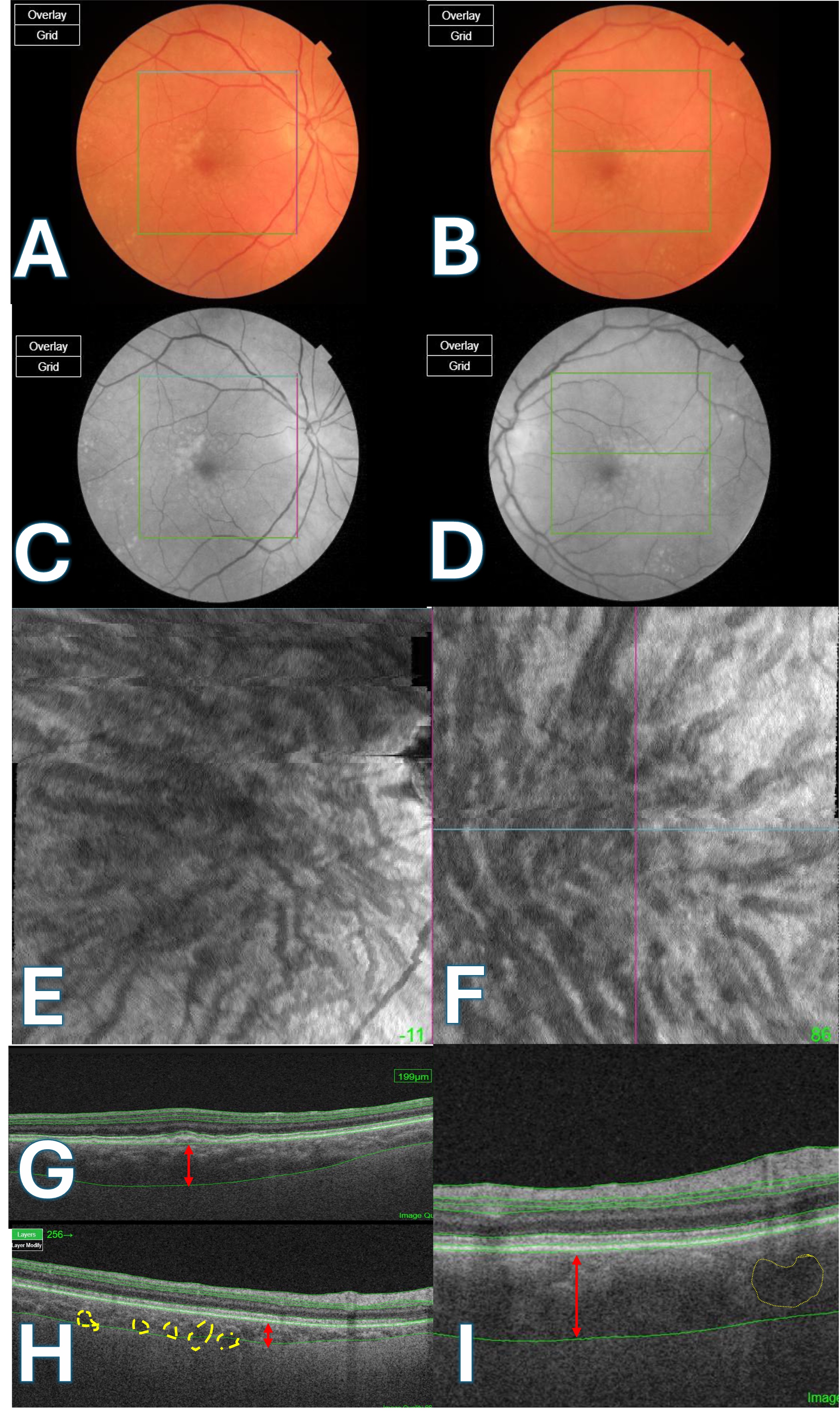 | 2,369 × 3,978 (1.43 MB) | Joobin.Khadamy (talk | contribs) |
You cannot overwrite this file.
File usage
The following 2 pages use this file: