File:Nipah Virus MRI Findings.jpg
From EyeWiki
Size of this preview: 800 × 283 pixels. Other resolution: 1,800 × 637 pixels.
Original file (1,800 × 637 pixels, file size: 97 KB, MIME type: image/jpeg)
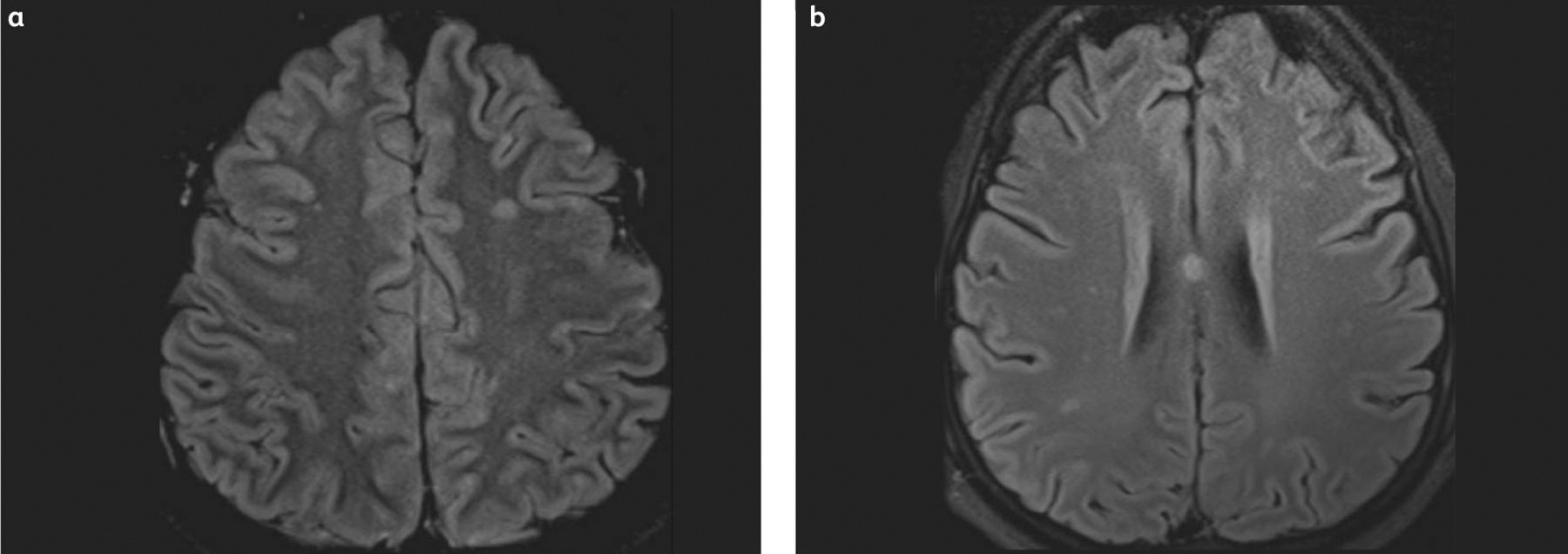
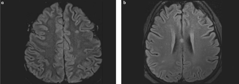
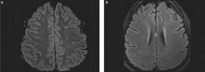
Figure 2: (a) Magnetic resonance imaging using fluid-attenuated inversion recovery shows multiple small bright lesions with areas of cortical involvement. (b) Magnetic resonance imaging using diffusion-weighted imaging demonstrates multiple bright lesions on both sides of the brain. Open access under terms of the Creative Commons License 4.0: https://creativecommons.org/licenses/by-nc-nd/4.0/. Original figures available at: https://doi.org/10.7861/clinmed.2022-0166
File history
Click on a date/time to view the file as it appeared at that time.
| Date/Time | Thumbnail | Dimensions | User | Comment | |
|---|---|---|---|---|---|
| current | 19:42, December 5, 2025 | 1,800 × 637 (97 KB) | Dina.Abdelsalam (talk | contribs) | ||
| 19:41, December 5, 2025 | 678 × 240 (26 KB) | Dina.Abdelsalam (talk | contribs) |
You cannot overwrite this file.
File usage
The following page uses this file: