.jpg)
.jpg&action=edit&redlink=1)
.jpg&action=edit)
.jpg&action=history)
File:Scleral Buckle Placement (Encircling Element in Surgery).jpg
Original file (986 × 658 pixels, file size: 120 KB, MIME type: image/jpeg)
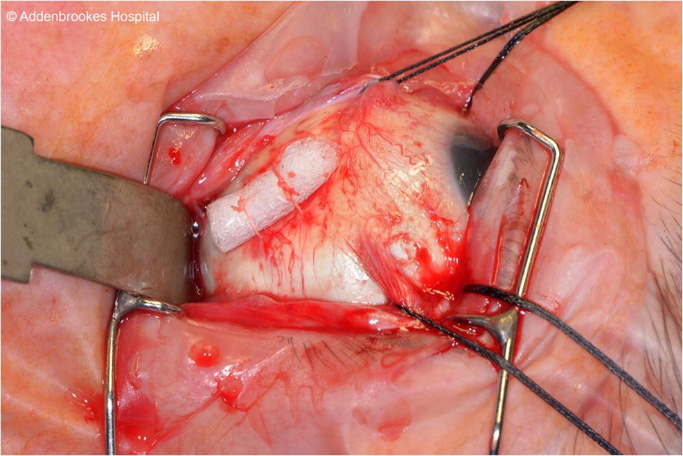
Intraoperative photograph demonstrating placement of an encircling scleral buckle beneath the extraocular muscles during retinal detachment repair. The silicone explant is secured to the scleral surface, with muscle hooks and traction sutures providing exposure. This image illustrates key anatomic relationships and surgical steps in modern scleral buckling technique. Image © Addenbrooke’s Hospital. Source: Graefe’s Archive for Clinical and Experimental Ophthalmology, “Scleral buckling—a brief historical overview and current status,” available at https://link.springer.com/article/10.1007/s00417-019-04562-1.
File history
Click on a date/time to view the file as it appeared at that time.
| Date/Time | Thumbnail | Dimensions | User | Comment | |
|---|---|---|---|---|---|
| current | 14:19, December 11, 2025 | | 986 × 658 (120 KB) | David.TaylorGonzalez (talk | contribs) |
You cannot overwrite this file.
File usage
The following page uses this file:
.jpg&oldid=123775)