File:Stingray MRI.jpeg
Stingray_MRI.jpeg (800 × 398 pixels, file size: 69 KB, MIME type: image/jpeg)
Summary
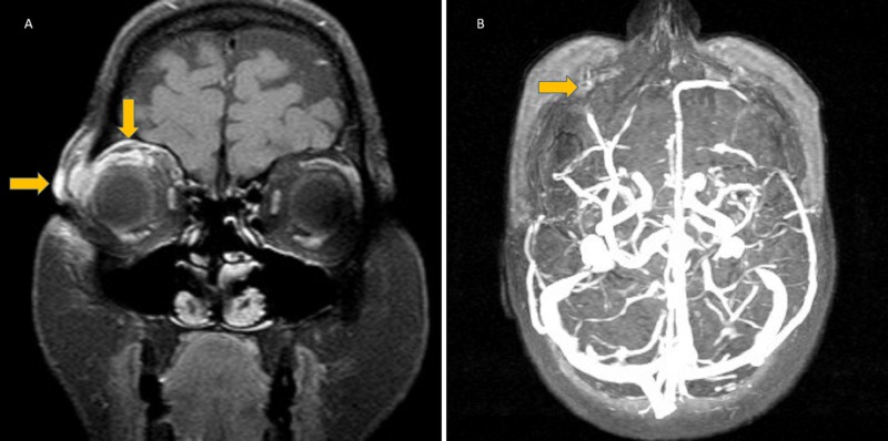
Figure 5. Stingray tail ocular injury. A) T1 SPIR contrast enhancement on a coronal MRI slice displaying right eye subcutaneous edema and periorbital cellulitis. (B) Thrombosis of right eye superior ophthalmic vein.[1]
Copyright © 2020, Ozunal et al.
This is an open access article distributed under the terms of the Creative Commons Attribution License, which permits unrestricted use, distribution, and reproduction in any medium, provided the original author and source are credited.
As a library, NLM provides access to scientific literature. Inclusion in an NLM database does not imply endorsement of, or agreement with, the contents by NLM or the National Institutes of Health.
Learn more: PMC Disclaimer | PMC Copyright Notice
- ↑ Ozunal ZG, Karsidag S, Sahin S. Superior Optic Vein Thrombosis Related to Orbital Cellulitis Secondary to Aquatic Injury. Cureus. 2020;12(2):e7080. doi:10.7759/cureus.7080
File history
Click on a date/time to view the file as it appeared at that time.
| Date/Time | Thumbnail | Dimensions | User | Comment | |
|---|---|---|---|---|---|
| current | 18:25, December 23, 2025 | | 800 × 398 (69 KB) | Talhah.Zubair (talk | contribs) | Figure 5. Stingray tail ocular injury. A) T1 SPIR contrast enhancement on a coronal MRI slice displaying right eye subcutaneous edema and periorbital cellulitis. (B) Thrombosis of right eye superior ophthalmic vein.38 Copyright © 2020, Ozunal et al. This is an open access article distributed under the terms of the Creative Commons Attribution License, which permits unrestricted use, distribution, and reproduction in any medium, provided the original author and source are credited. As a libra... |
You cannot overwrite this file.
File usage
The following page uses this file: