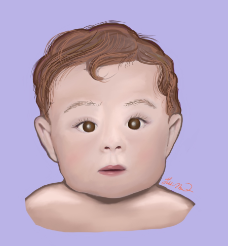
Galactokinase Deficiency
All content on Eyewiki is protected by copyright law and the Terms of Service. This content may not be reproduced, copied, or put into any artificial intelligence program, including large language and generative AI models, without permission from the Academy.
Disease Entity
Disease
Galactokinase deficiency, commonly referred to as Type II Galactosemia, is one of four recognized inborn errors of galactose metabolism—also referred to as galactosemias. This autosomal recessive mutation of the galactokinase enzyme results in the accumulation of galactitol, a metabolite associated with galactose metabolism. The most common ophthalmic manifestation of the disease is the development of bilateral nuclear sclerotic cataracts.
Epidemiology
Galactokinase deficiency is one of the mildest forms of galactosemia in terms of systemic disease. The prevalence of this condition ranges from 1 in 50,00 to 2,200,000.[1] In the United States the estimated incidence of galactokinase deficiency is 1 in 100,000 neonates.[1]
Etiology
Galactokinase deficiency results from a mutation in the galactokinase enzyme (GALK). The galactokinase enzyme is encoded two genes: the GALK1 gene located on chromosome 17q25.1,[2] and the GALK2 gene located on chromosome 15q21.1-q21.2.[3] Notably, galactokinase deficiency is associated with GALK1 mutations, for which over 30 mutations have been identified to date.[1]
Pathophysiology
An in-depth explanation of the pathophysiology behind galactosemias is explained elsewhere. Briefly, galactose is primarily metabolized by the Leloir pathway. However, when enzymes, such as galactokinase are deficient, metabolites are shunted to alternative pathways—the most clinically-relevant of which being the aldolase reductase pathway. This alternative pathway is responsible for the production of a metabolite known as galactitol, which has osmotic properties.
Due to the increased presence of aldolase reductase on the anterior surface of the lens, increased levels of galactose in the body in the presence of a mutated galactokinase enzyme cause the lens to accumulate increased galactitol levels. This accumulation can lead to lens swelling, cell lysis, and ultimately cataract formation.[4]
Diagnosis
Physical Exam
Galactokinase deficiency can be difficult to recognize in the early stages of life due to the lack of dramatic symptomatic manifestations that present. The most common ophthalmic manifestation of the disease is the development of bilateral nuclear sclerotic cataracts which can sometimes go unrecognized, leading to nystagmus, failure to develop social smile, or diminished ability to track objects.[1] In addition to causing galactosemia in individuals with homozygous mutations, heterozygous carriers are at an increased risk of pre-senile cataracts (earlier than 40 years of age).[5]
Systemically, hyperbilirubinemia is one of the more common signs which presents in the neonatal period. Pseudotumor cerebri (also known as Idiopathic Intracranial Hypertension) can be due to increased cerebrospinal fluid from osmotic pressure caused by the increased galactitol concentration. Long term follow-up of patients with galactosemia has also revealed a higher risk of bleeding diathesis, encephalopathy, dyspraxia, mental retardation, motor delays, and hypergonadotropic hypogonadism.[1]
Differential Diagnosis
When evaluating a patient with suspected galactokinase deficiency, it is important to consider all variants of inborn errors of galactose metabolism as part of the differential diagnosis. In addition, it is necessary to consider other conditions that can cause cataracts at the individual’s age.
Management
General treatment
Early detection by newborn bloodspot screening and dietary modifications are essential to achieving favorable outcomes.[6] Dietary restriction has been associated with a decreased incidence of cataract formation and an increased likelihood of resolution for those who have already developed cataracts.[6] When treatment is started within two to three weeks of age, reversal of cataract formation can occur.[1] In particular, it is recommended that patients have strict galactose restriction with calcium supplementation to prevent the accumulation of galactitol.[6] In infants, a soy-based formula is recommended.
Despite the introduction of a galactose-restricted diet, there is still a possibility of developing cataracts; however, it is uncertain whether this is due to lack of adherence to the diet or an alternative pathological mechanism.[6]
Follow - Up
Long-term follow-up with the patients’ primary care provider is necessary to monitor their measurements of galactose, its derivative metabolites, and measurements of galactitol in red blood cells.[1] Routine ophthalmologic evaluations are also necessary to monitor for cataract formation. If a patient develops a visually significant cataract, cataract surgery will be required with or without intraocular lens implantation.
References
- ↑ 1.0 1.1 1.2 1.3 1.4 1.5 1.6 Ramani PK, Arya K. Galactokinase Deficiency. [Updated 2022 Mar 7]. In: StatPearls [Internet]. Treasure Island (FL): StatPearls Publishing; 2022 Jan-. Available from: [1]
- ↑ Elsevier SM, Kucherlapati RS, Nichols EA, et al. Assignment of the gene for galactokinase to human chromosome 17 and its regional localisation to band q21-22. Nature. 1974;251(5476):633-636. doi:10.1038/251633a0
- ↑ Lee RT, Peterson CL, Calman AF, Herskowitz I, O’Donnell JJ. Cloning of a human galactokinase gene (GK2) on chromosome 15 by complementation in yeast. Proc Natl Acad Sci U S A. 1992;89(22):10887-10891. doi:10.1073/pnas.89.22.10887
- ↑ Ai Y, Zheng Z, O’Brien-Jenkins A, et al. A mouse model of galactose-induced cataracts. Hum Mol Genet. 2000;9(12):1821-1827. doi:10.1093/hmg/9.12.1821
- ↑ Skalka HW, Prchal JT. Presenile Cataract Formation and Decreased Activity of Galactosemic Enzymes. Arch Ophthalmol. 1980;98(2):269-273. doi:10.1001/archopht.1980.01020030265003
- ↑ 6.0 6.1 6.2 6.3 Rubio-Gozalbo ME, Derks B, Das AM, et al. Galactokinase deficiency: lessons from the GalNet registry. Genet Med Off J Am Coll Med Genet. 2021;23(1):202-210. doi:10.1038/s41436-020-00942-9


