Corneal Dellen
All content on Eyewiki is protected by copyright law and the Terms of Service. This content may not be reproduced, copied, or put into any artificial intelligence program, including large language and generative AI models, without permission from the Academy.
Corneal dellen are peripheral excavations in the cornea occurring secondary to tear film disturbance caused by limbal elevations.
Disease Entity
ICD-10: H18.49
Disease
“Dellen” dimple was first described by Ernst Fuchs as shallow, saucer-like excavations at the margin of the cornea. [1]
They are thought to occur due to localized tear film instability especially the mucin layer and dehydration. [2]
Etiology
- Secondary to paralimbal elevation due to [1][3]
- Episcleritis or scleritis
- Thick pingecula or pterygium
- Subconjunctival hemorrhage
- Severe conjunctival chemosis
- Subconjunctival injections
- Filtering blebs
- Suture granuloma
- Limbal tumours
- Lesions like angioma
- Subconjunctival silicone oil
- Post-surgery – cataract/rectus muscle surgery/trabeculectomy [4][5]
- Long term contact lens wear [6]
- Following paralytic lagophthalmos
- Idiopathic, in elderly persons
- Ocular trauma
Pathophysiology
- Paralimbal elevation causes localised break in the tear film, especially a focal absence of the mucin layer.
- Cornea epithelium is hydrophobic and in absence of mucin will repel water causing a dry spot and eventually localized dehydration.
Diagnosis
History
History of:
- Ocular surgery
- Ocular trauma
- Previous ocular complaints
- Contact lens wear
Physical Examination
Symptoms
- Redness
- Foreign body sensation
- Grittiness
Signs
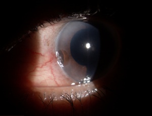
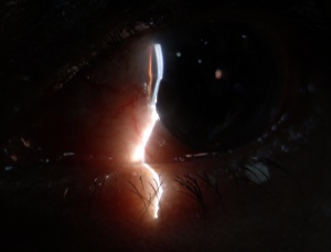
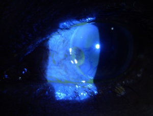
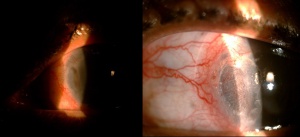
Slit lamp examination reveals [1]
- Depressed region with clearly defined margins
- Around 2-3 mm in diameter
- Commonly on the temporal side, forming ellipses parallel to the limbus
- Corneal wall is steep, limbal wall is sloping
- Epithelium is usually intact overlying a thinned area of dehydrated stroma
- Fluorescein pools in the depression and at times there is overlying staining
- Epithelium within the dellen can either be clear or opaque (hazy and dry) in appearance
- Corneal sensation can be decreased with the dellen compared to rest of the cornea
- Adjoining vascular loops and conjunctival vessels can be injected
- Adjacent cornea is usually normal
Progression
- Dellens usually last for 24-48 hours and disappear spontaneously. Most will heal within 2 weeks.[8]
- However, they can become chronic with breakdown of epithelium, inflammation of stroma and scarring.
Complications
- If left untreated, the underlying corneal stroma may undergo secondary degeneration, leading to corneal scarring and/or vascularisation with accompanying loss of vision. [9]
- Rarely can lead to corneal perforation. [10]
Management
Treatment needs to be initiated expeditiously to avoid the above mentioned complications.
General treatment
- Reduction of the paralimbal elevation i.e. treating the underlying cause
Medical therapy
- Rapid re-establishment of the mucin layer and a hydrophilic corneal surface by:
- Frequent lubrication with artificial tears and ointments
- Patching the eye
- Large diameter bandage contact lens[11]
- Dellens following filtering blebs – limbal elevation cannot be changed in these cases as bleb is necessary. These patients benefit with chronic frequent use of artificial tears.
Surgery
Surgical excision of paralimbal elevations like pterygium, pingecula, limbal tumours etc.
Minor procedures like tarsorraphy to limit ocular surface exposure can be considered.
Additional Resources
- Boyd K, Pagan-Duran B, Hazanchuk V. Lubricating Eye Drops. American Academy of Ophthalmology. EyeSmart/Eye health. https://www.aao.org/eye-health/treatments/lubricating-eye-drops-2. Accessed February 28, 2023.
References
- ↑ 1.0 1.1 1.2 Fuchs, Adalbert. "Pathological dimples (“Dellen”) of the cornea." American Journal of Ophthalmology 12.11 (1929): 877-883.
- ↑ Mai G, Yang S. Relationship between corneal dellen and tearfilm breakup time. Yan Ke Xue Bao 1991;7(1):43-6.
- ↑ 3.0 3.1 Accorinti M, Gilardi M,Giubilei M, De Geronimo D, Iannetti L. Corneal and scleral dellen after an uneventful pterygium surgery and a febrile episode. Case Rep Ophthalmol. 2014;5(1):111‐115.
- ↑ Fresina M, Campos E. Corneal “dellen” as a complication of strabismus surgery. Eye (Lond) 2009;23(1):161-3.
- ↑ Jampel, Henry D., et al. "Perioperative complications of trabeculectomy in the collaborative initial glaucoma treatment study (CIGTS)." American journal of ophthalmology 140.1 (2005): 16-22.
- ↑ Ng LH. Central corneal epitheliopathy in a long term, overnight orthokeratology lens wearer: a case report. Optom Vis Sci 2006;83(10):709-14.
- ↑ Baum, Jules L., Saiichi Mishima, and S. Arthur Boruchoff. "On the nature of dellen." Archives of Ophthalmology 79.6 (1968): 657-662.
- ↑ Corneal 'dellen' as a complication of strabismus surgery. Fresina M, Campos ECEye (Lond). 2009 Jan; 23(1):161-3.
- ↑ Dua HS, Forrester JV. Clinical patterns of corneal epithelial wound healing. Am J Ophthalmol 1987;104:481
- ↑ González Gomez A, Dellen and corneal perforation after bilateral pterygium excision in a patient with no risk factors. BMJ Case Rep. 2015 Nov 30;2015
- ↑ Kymionis, George D., et al. "Treatment of corneal dellen with a large diameter soft contact lens." Contact Lens and Anterior Eye 34.6 (2011): 290-292.


