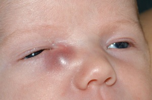
Dacryocele
All content on Eyewiki is protected by copyright law and the Terms of Service. This content may not be reproduced, copied, or put into any artificial intelligence program, including large language and generative AI models, without permission from the Academy.
Disease Entity
ICD10: Q10.5
Disease
Dacryocele is also known as a dacryocystocele, amniotocele, amniocele, or mucocele. It is formed when a distal blockage (usually membranous) of the lacrimal sac causes distention of the sac, which also kinks and closes off the entrance to the common canaliculus. This restricts retrograde discharge of the accumulated secretions and prevents decompression of the sac. Dacryoceles may present in 3% of infants with NLDO and are most commonly unilateral, but can be bilateral in up to 30% of patients.[1]
Etiology
A dacryocele forms within the lacrimal sac +/- nasal cavity as a result of a congenital nasolacrimal duct obstruction (NLDO). The mucus secreted by the lacrimal sac goblet cells or amniotic fluid becomes trapped in the nasolacrimal sac secondary to a distal blockage. The trapping of the fluid then causes distention of the sac, which can close off the entrance from the common canaliculus The valve of Rosenmüller acts as a one-way valve restricting retrograde discharge of the accumulated secretions preventing decompression of the sac.[2]
Risk Factors
Nasolacrimal duct obstruction
Diagnosis
History
Patients are usually infants who present at birth or within the first few days of life with a bluish swelling just below and nasal to the medial canthus. They also have a NLDO and may have mucopurulent discharge or mattering of lashes, most notable after awakening. If untreated it can progress to dacryocystitis, rupture and fistula formation or even airway blockage. [1] [2][3][4]
Physical Examination
Exam reveals a bluish, cystic mass that is located below the medial canthal tendon in the area of the nasolacrimal sac. The medial canthus can be displaced superiorly. Gentle pressure over the mass can produce mucopurulent discharge from the eyelid puncta. In the majority of cases, the nasal mucosa becomes distended and extend into the nasal cavity.[5] This nasal cystic extension may be visualized during endoscopic nasal examination under the inferior turbinate. [1] [2][3][4] Therefore, the nasal cavity should always be evaluated to assess for an intranasal cyst extension that may cause airway obstruction.
Signs
A cystic mass present inferior to the medial canthus with a bluish discoloration of the overlying skin. Mucopurulent discharge or mattering of lashes, most notable after awakening. Mucopurulent discharge on digital palpation. [1][2][3][4]
Symptoms
Mucopurulent discharge or mattering of lashes, most notable after awakening. Historically, it has been stated that infants are obligate nasal breathers. However, further studies have demonstrated that infants can breathe through their mouths, if necessary, although nasal breathing is highly preferred as it allows them to feed at the same time. In severe cases with large intranasal cysts or bilateral obstructions, infants may present with respiratory symptoms of airway obstruction ranging from breathing difficulty when feeding to acute respiratory distress. [1][2][3][4]
Clinical Diagnosis
Diagnosis is made with clinical examination. It can be helpful to perform digital massage to attempt expression of fluid from the area. Nasal examination is warranted to evaluate for an intranasal cyst extension that may affect breathing. [1][2][3][4]
Diagnostic Procedures
It is a clinical diagnosis, however due to frequent association with endonasal cysts, an endoscopic nasal examination is often warranted. Although imaging is not often warranted, in cases of endonasal cysts, computed tomography scan or magnetic resonance imaging will show a large cystic mass extending from the lacrimal system into the inferior meatus. Ultrasonography can also be used in the detection of a dacryocele. B-scan will reveal a hollow round cavity with an ostium connected with the nasolacrimal duct, and the A-scan will reveal the high reflecting walls and very low internal reflectivity. [6]
Laboratory Tests
No laboratory tests are necessary
Differential diagnosis
The differential diagnosis includes hemangioma, dermoid cyst, encephalocele, rhabdomyosarcoma, and other solid tumors of the lacrimal system, including pylomatrixoma[1]. Imaging including CT and MRI scans can help differentiate a dacryocele from the others that are listed below with some differentiating clinical characteristics:
- Hemangioma - has irregular borders and inhomogeneous consistency, will not be present at birth, will increase in size with head down position, and is less firm when compared to a dacryocele.
- Dermoid cyst- well delineated, mobile, skin colored cyst located above the medial canthal tendon
- Encephalocele- located above the medial canthal tendon
- Rhabdomyosarcoma- rapidly progressing mass, not present at birth
Management
Prompt treatment of dacryocystocele is recommended, even in the absence of respiratory symptoms, to minimize the risk of complications, particularly infections. Given that infants have underdeveloped immune systems, they are more vulnerable to both localized and systemic infections. Since dacryocystoceles are initially sterile, conservative management with gentle digital massage of the lacrimal sac combined with prophylactic antibiotics may be effective in resolving the condition.
However, if there is no improvement within the first one to two weeks of life, or if signs of infection (acute dacryocystitis) develop, surgical intervention becomes necessary. Nasolacrimal duct (NLD) probing alone may be sufficient in some cases, though about 25% of infants may not achieve complete resolution.[3][7] For these patients, combining NLD probing with nasal endoscopy and marsupialization of the intranasal cyst significantly improves outcomes, with success rates reaching >95%.[7]
In cases of infection, systemic antibiotics should be administered preoperatively and surgery performed once the infection has subsided. Importantly, drainage of an infected dacryocele through a skin incision is discouraged, as it may lead to the formation of a persistent fistula.[1]
If an infant presents with acute respiratory distress due to obstruction from a nasal cyst associated with a dacryocystocele, urgent nasal endoscopy and elimination of the cyst is indicated. This can be performed at the bedside by a pediatric ENT specialist and in most cases resolves the blockage and prevents recurrences.
Complications
If the dacryocele does not resolve with conservative management, infection may develop within the first few weeks of life. In acute dacryocystitis, the skin over the lacrimal sac becomes erythematous and pressure over the sac may produce purulent discharge. Infections require management with systemic antibiotics. If improvement does not occur, probing of the nasolacrimal system as well as marsupialization of the nasal mucocele are needed to decompress the sac and allow the externalization of the contents.[1][3] Infants with acute respiratory distress require immediate surgical management.[1]
Prognosis
The prognosis is excellent
References
- ↑ 1.0 1.1 1.2 1.3 1.4 1.5 1.6 1.7 1.8 1.9 Basic Clinical and Science Course. Pediatric Ophthalmology and Strabismus. 2025-26. Chapter 18 pg 246-248.
- ↑ 2.0 2.1 2.2 2.3 2.4 2.5 Basic Clinical and Science Course. Orbit, Eyelids, and Lacrimal System. 2011-12. Section 7 pg 272.
- ↑ 3.0 3.1 3.2 3.3 3.4 3.5 3.6 Becker BB. The treatment of congenital dacryocystocele. Am J Ophthalmology. 2006; 142(5):835- 838.
- ↑ 4.0 4.1 4.2 4.3 4.4 Yen, KG; Yen, MT. “Initial Management of the Tearing Infant.” EyeNet July 2004. http://www.aao.org/publications/eyenet/200407/pearls.cfm?RenderForPrint=1&
- ↑ Lueder G.T.: The association of neonatal dacryocystoceles and infantile dacryocystitis with nasolacrimal duct cysts. Trans Am Ophthalmol Soc 2012; 110: pp. 74-93.
- ↑ Cavazza, S, et al. “Congenital Dacryocystocele: Diagnosis and Treatment. “ Acta Otorhinolaryngol Ital. 2008 December; 28(6): 298–301.
- ↑ 7.0 7.1 Ahmad TR, Lindquist KJ, Oatts JT, de Alba Campomanes AG, Kersten RC, Chan DK, Indaram M. Probing versus primary nasal endoscopy for the treatment of congenital dacryocystoceles. J AAPOS. 2024 Apr;28(2):103865. doi: 10.1016/j.jaapos.2024.103865. Epub 2024 Mar 6. PMID: 38458602.


